Numerous collagen stimulation treatments have been extensively researched over the past two decades. Unlike traditional fillers that restore volume, these treatments target the underlying extracellular matrix, which activates fibroblasts and initiates sustained collagen synthesis.
This article provides a comprehensive clinical overview of the most evidence-based collagen-inducing therapies, including:
- Poly-L-lactic acid and calcium hydroxylapatite-based collagen-stimulating injections
- Microfocused ultrasound with real-time visualization
- Microneedling
- Fractional laser
- Radiofrequency
Let’s get started.
Key Takeaways
- Sculptra® (PLLA) and Radiesse® (CaHA) are FDA-approved injectables with well-documented neocollagenesis through fibroblast-mediated type I collagen deposition.
- Ultherapy targets dermal and SMAS layers at depths of 1.5–4.5 mm, producing thermal injury zones (~1 mm³) that stimulate type I/III collagen and elastin.
- Microneedling triggers controlled dermal trauma, activating TGF-β, PDGF, and VEGF pathways. Split-face trials confirm increased collagen types I, III, and VII, with clinical improvements comparable to ablative lasers.
- Ablative (CO₂, Er: YAG) and non-ablative fractional lasers (1540–1550 nm) improve ECM turnover through thermal coagulation or ablation.
- Non-ablative lasers are preferred in Fitzpatrick IV–VI due to the lower risk of post-inflammatory hyperpigmentation.
- Radiofrequency induces dermal collagen denaturation and subsequent neocollagenesis via TGF-β and HSP47 signaling.
5 Collagen Producing Treatments With Strong Clinical Evidence
Here is the clinically grounded update on the five best collagen-producing treatments with proven efficacy and favorable safety profiles.
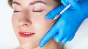
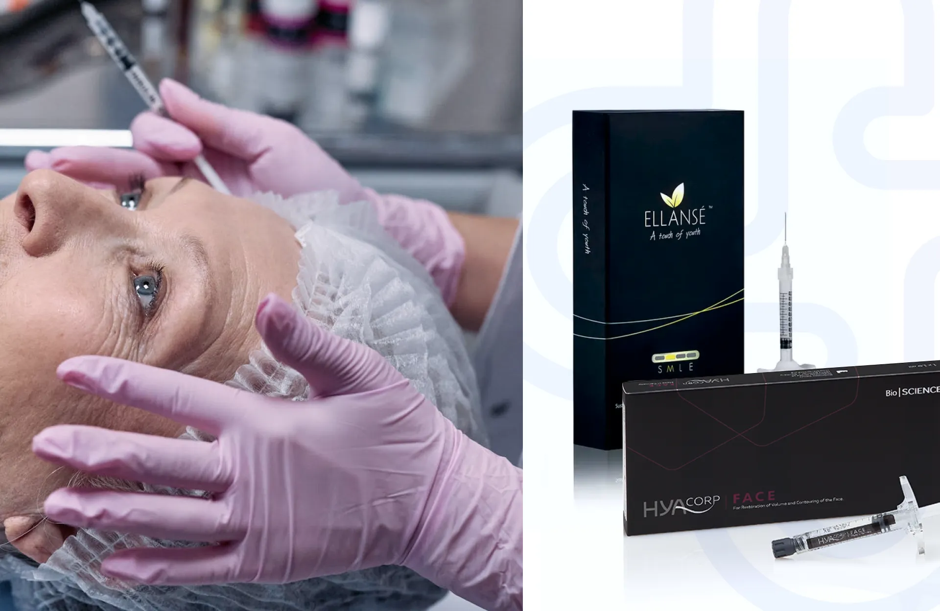
1. Biostimulator Injections
Biostimulator injections play the dual role of volumization and neocollagenesis. Unlike hyaluronic acid fillers, these collagen-stimulating fillers induce controlled fibroplasia for progressive dermal remodeling and long-term structural improvements.
Among the FDA-approved biostimulatory agents, Sculptra® (poly-L-lactic acid) and Radiesse® (calcium hydroxylapatite) have the most robust body of evidence.
Sculptra® (Poly-L-lactic acid)
Sculptra® (PLLA) is a synthetic, biodegradable polymer filler that produces collagen via a subclinical inflammatory response. PLLA microparticles stimulate fibroblasts through a foreign-body reaction, leading to type I collagen deposition over a 6–12 month period post-injection.
A 2004 randomized controlled trial showed significant improvement in facial volume restoration through Sculptra in HIV-associated lipoatrophy, with histological confirmation of new collagen formation.1
Adverse effects of Sculptra collagen stimulation are largely technique-dependent. When following the updated injection protocols (e.g., dilution with 5–8 mL sterile water and deep dermal or subcutaneous administration), the incidence of complications is significantly reduced.2
Radiesse® (Calcium Hydroxylapatite)
Radiesse® consists of 25–45 µm calcium hydroxyapatite (CaHa) microspheres suspended in a carboxymethylcellulose (CMC) gel carrier. The CaHA acts as a scaffold for fibroblast infiltration and subsequent collagen synthesis, while the CMC provides immediate volumization.
In a histological study by Berlin et al. (2008), biopsies taken at 3, 6, and 12 months post-Radiesse collagen stimulation injection showed progressive collagen type I deposition replacing the gel carrier.3
Emerging uses of Radiesse in regenerative medicine, including hand rejuvenation and skin laxity improvement, have also been validated.
Radiesse is best suited for deep dermal or supraperiosteal injection, and newer protocols include hyperdilution techniques (1:1 to 1:4 with saline/lidocaine).
The side effects of CaHa fillers are limited to transient edema or bruising. Vascular occlusion risk is lower than with HA due to the product’s higher viscosity and its non-hydrophilic nature.
2. Ultherapy
Ultherapy® is an FDA-cleared non-invasive treatment that uses micro-focused ultrasound with real-time visualization (MFU-V).
Ultherapy induces a precisely targeted thermal injury (60–70°C) to the reticular dermis and superficial musculoaponeurotic system at pre-defined depths (1.5 mm, 3.0 mm, and 4.5 mm).
These precise thermal injury zones (TIZs) are approximately 1 mm³ in volume. They trigger immediate collagen denaturation and contraction, which in turn initiate long-term neocollagenesis and neoelastogenesis.
The overall effect of Ultherapy is fibroblast activation, upregulation of heat shock proteins, and subsequent type I and III collagen synthesis, as well as tropoelastin expression.
Multiple clinical trials have validated Ultherapy as one of the best collagen-boosting treatments. For example, Marquardt et al. used micro-focused ultrasound and confirmed increased dermal fibroblasts and mature collagen, and newly synthesized elastin three months following treatment.4
Ultherapy’s results are cumulative, with optimal collagen induction seen at 3–6 months. The real-time visualization reduces the risk of off-target heating, and no permanent nerve injury or scarring has been reported in large multicenter safety reviews so far.
3. Microneedling
Microneedling, also referred to as percutaneous collagen induction (PCI), is a minimally invasive procedure that uses sterile, fine-gauge needles to create controlled micro-injuries within the epidermis and dermis. It has established utility in aesthetic dermatology and regenerative medicine for the treatment of rhytides, acne scarring, skin laxity, and post-inflammatory dyschromia.
Microneedling induces punctate dermal trauma, which activates keratinocytes, fibroblasts, and platelets. It stimulates the release of TGF-β, PDGF, and VEGF, all of which cause an increased synthesis of type I and III collagen, fibronectin, and elastin fibers within the extracellular matrix.
El-Domyati et al. conducted a split-face comparative study assessing microneedling vs. CO₂ laser in patients with photoaged skin. Biopsies taken post-treatment confirmed increased collagen types I, III, and VII on the microneedled side. Improvements in dermal density and texture were also reported to be comparable to ablative laser resurfacing but with fewer adverse effects.5
Microneedling is also well tolerated across all Fitzpatrick skin types due to its non-ablative nature and preservation of the epidermal barrier.6
In addition, the use of FDA-approved microneedling pens with disposable cartridges mitigates the risk of infection or cross-contamination. Adjunctive therapies (e.g., PRP, vitamin C, and exosomes) have also been studied in the literature, but they must be scrutinized for patient compatibility and immunogenicity prior to percutaneous delivery.
4. Laser for Collagen Stimulation
Laser-based collagen stimulation is also widely recognized. The most clinically validated wavelengths for collagen induction include CO₂ (10,600 nm), Er: YAG (2940 nm), and non-ablative fractional devices (e.g., 1540–1550 nm erbium-glass lasers).7
Thermal injury from fractional lasers initiates a controlled wound-healing cascade, involving cytokine release, heat-shock protein activation, and fibroblast recruitment. These processes collectively stimulate the synthesis of type I and III collagen and dermal matrix glycoproteins.
Ablative laser for collagen stimulation vaporizes microcolumns of tissue (microthermal treatment zones), while non-ablative devices induce sub-epidermal coagulation without surface ablation.
Magni et al. assessed baseline collagen expression in human dermal fibroblasts (HDFa) by measuring fluorescence intensity without laser exposure. Treatment with a 1540-nm fractional laser at energy densities of 3.5 and 2.8 J/cm² led to a significant upregulation in fluorescence intensity corresponding to type III collagen in human dermal fibroblasts.8
Ablative lasers (e.g., CO₂ and Er: YAG) carry higher risks of post-inflammatory hyperpigmentation, especially in Fitzpatrick IV–VI skin types. However, non-ablative fractional systems have a favorable downtime profile with lower epidermal disruption.
5. Radiofrequency Collagen Stimulation
Radiofrequency (RF)-based therapies are widely established in non-ablative skin rejuvenation protocols.
RF generates an oscillating electrical current that induces ionic agitation and frictional heating within the reticular dermis and subdermal tissues.
RF energy, delivered in monopolar, bipolar, or multipolar configurations, produces volumetric dermal heating in the range of 40–45°C.
The thermal injury denatures collagen triple helices and initiates a cascade of molecular events involving TGF-β, HSP47, and IL-6. As a result, fibroblast stimulation occurs with upregulation of type I and III collagen synthesis.
Uribe et al. observed enhanced basophilic staining indicative of thin type I and III collagen fibers in 12 skin biopsies collected from six patients in a study. In parallel, molecular assays demonstrated elevated fibroblast activity, attributed to the upregulation of heat shock protein HSP47.9
Radiofrequency collagen stimulation also has a favorable safety profile across all skin phototypes due to its non-chromophore-dependent mechanism.
Source High-Quality Collagen-Stimulators at the Best Prices From Medica Depot
As clinical demand for collagen stimulators continues to grow, ensuring product authenticity and consistency is important for practitioners aiming to deliver safe, evidence-based care.
Medica Depot is a trusted global distributor offering a comprehensive range of high-quality, collagen-stimulating biostimulators and dermal fillers sourced directly from manufacturers. We empower licensed medical professionals to procure the best-in-class products at competitive wholesale pricing.
Visit Medica Depot today to explore our full catalog.
FAQs
Are Collagen-Stimulating Injections Safe?
When administered using proper technique and dilution protocols, collagen-stimulating injectables such as PLLA and CaHA demonstrate a favorable safety profile. Mild adverse reactions are reported for most treatments, but they go away in a few days.
How Long Does Collagen Stimulation Last?
Collagen-stimulating treatments start working within weeks and continue for several months post-treatment. Clinical effects from biostimulators persist for 6–24 months, depending on agent type, anatomical site, and individual tissue response.
What Is the Difference Between Collagen Stimulator and Filler?
Collagen stimulators induce endogenous neocollagenesis through controlled tissue activation. Hyaluronic acid-based fillers, in contrast, provide immediate volume replacement without stimulating new collagen synthesis.
References
1. Vleggaar D, Bauer U. Facial enhancement and the European experience with Sculptra (poly-l-lactic acid). Journal of drugs in dermatology: JDD. 2004;3(5):542-547. https://pubmed.ncbi.nlm.nih.gov/15552606/
2. SCULPTRA ® Poly-L-Lactic Acid. Accessed July 13, 2025. https://www.galderma.com/sites/default/files/2025-03/IFU_Sculptra-Jul_2022.pdf
3. BERLIN AL, HUSSAIN M, GOLDBERG DJ. Calcium Hydroxylapatite Filler for Facial Rejuvenation: A Histologic and Immunohistochemical Analysis. Dermatologic Surgery. 2008;34(s1):S64-S67. doi:https://doi.org/10.1111/j.1524-4725.2008.34245.x
4. Marquardt K, Hartmann C, Wegener F, et al. Microfocused Ultrasound With Visualization Induces Remodeling of Collagen and Elastin Within the Skin. Journal of Cosmetic Dermatology. Published online November 15, 2024. doi:https://doi.org/10.1111/jocd.16638
5. El-Domyati M, Barakat M, Awad S, Medhat W, El-Fakahany H, Farag H. Microneedling Therapy for Atrophic Acne Scars: An Objective Evaluation. The Journal of Clinical and Aesthetic Dermatology. 2015;8(7):36-42. https://pubmed.ncbi.nlm.nih.gov/26203319/
6. STL Volume 21 Number 1. Melasma and Post Inflammatory Hyperpigmentation Treatment Update. Skin Therapy Letter. Published 2016. https://www.skintherapyletter.com/hyperpigmentation/melasma-post-inflammatory-hyperpigmentation-treatment/
7. Heidari Beigvand H, Razzaghi M, Rostami-Nejad M, et al. Assessment of Laser Effects on Skin Rejuvenation. Journal of Lasers in Medical Sciences. 2020;11(2):212-219. doi:https://doi.org/10.34172/jlms.2020.35
8. Magni G, Piccolo D, Bonan P, et al. 1540-nm fractional laser treatment modulates proliferation and neocollagenesis in cultured human dermal fibroblasts. Frontiers in Medicine. 2022;9. doi:https://doi.org/10.3389/fmed.2022.1010878
9. Cala LC, Perez ME, Andreina Zannin Ferrero, et al. Effects of Bipolar Radiofrequency on Collagen Synthesis from Patients with Brachial Ptosis. Plastic and reconstructive surgery Global open. 2023;11(4):e4924-e4924. doi:https://doi.org/10.1097/gox.0000000000004924